Flower
The flower is concerned with the function of sexual reproduction in angiosperms.

Development of Male Gametophyte or Pollen
Stamen forms the male reproductive unit and is made up of anther and the filament. The anther is generally bi lobed with chamber containing several pollen grains.
Pollen grains are produced in the anther. A very young anther comprises a mass of undifferentiated thin - walled cells bounded by an epidermis. The anther becomes four lobed as the anther grows, and the lobes are joined by a sterile tissue called connective. Each lobe contains an elongated chamber which is termed as microsporangium or pollen sac.

(A) T.S. of young anther showing four microsporangia and a vascular stand;
(B) Detailed structure of one microsporangium;
(C) T.S. of mature anther
A cross section of a young anther reveals that the microsporangium is surrounded on the outside by a single layered epidermis. Beneath the epidermis is a layer of cells called the endothecium. The sporangial wall encloses a mass of cells characterised by their large size abundant cytoplasm and prominent nuclei. These cells are called sporogenous cells. These sporogenous cells undergo a few mitotic divisions to increase their number and form the microspore mother cells or microsporocytes. The microsporocytes are diploid and usually closely packed. Each diploid microsporocyte undergoes meiosis, giving rise to a group of four haploid cells, known as microspores or pollen grains. Such a group of 4 spores is known as a tetrad.

(A-C), Pollen grains or microspores.
(A-B), Entire pollen grains;
C, Pollen grain in section
At maturity, the partition wall between two adjacent microsporangia breaks down and the cavities of the two microsporangia merge into one pollen sac. When the pollen grains are fully formed, the ripe anther splits to liberate the pollen.
Structure of Pollen Grains
Each pollen grain is a haploid, unicellular mass of protoplast with a single nucleus. It is surrounded by a thick wall differentiated into two layers; the outer thick exine and the inner thin intine. The pollen grains reveal a wide range of microsculpturing of the exine under a scanning microscope. The exine is made up of a complex substance called sporopollenin. The pollen grains are the best preserved structures because the sporopollenin is one of the most resistant biological materials known. The size, form and ornamentation of the exine is quite characteristic of the plant and is of great taxonomic value.

The branch of science that deals with the study of the characteristics of the pollen grains is called palynology.
At one or more places, the exine is very thin or absent. These spots are called germ pores and are the points from which the pollen tubes emerge during pollen germination.
Development of Female Gametophyte
Pistil is the female reproductive unit, and is made up of a basal swollen part called the ovary, a stalk called the style and a terminal receptive disc called the stigma. The ovule begins its development as a minute protuberance on the placenta and forms a thick mass of cells termed as the nucellus. As the growth and development of the ovule proceeds, the nucellus is elevated on a short stalk like structure called the funiculus. The point at which the body of the ovule is attached to the funiculus is called as hilum. As the ovule develops further, one or two protective layers called the integuments grow from the base of the nucellus known as chalaza and surround the nucellus except for a narrow opening, the micropyle. This opening serves as a passage for the entry of a pollen tube into the ovule.


Development of embryo sac and female gamete (in an anatropous ovule)
A hypodermal cell of the nucellus enlarges and becomes differentiated into a megaspore mother cell or megasporocyte. This diploid megaspore mother cell increases in size and undergoes meiosis to form a linear tetrad of 4 haploid megaspores, 3 of which degenerate and the 4th becomes the functional megaspore.
Female Gametophyte
The nucleus of the megaspore undergoes three successive mitotic divisions forming eight nuclei. The megaspore enlarges into an oval shaped structure called the embryo sac. The eight nuclei of the embryo sac arrange themselves in 3 groups.


(A I), (A - D), stages in the development of an ovule and functional megaspore;
E I, stages in the development of an embryo sac from the functional megaspore
Three nuclei are towards the micropylar end, and form the egg apparatus with a central egg cell surrounded by 2 synergids. Three nuclei are at the chalazal end and form the antipodals. One nucleus from each pole moves to the central position of the embryo sac and are called as polar nuclei. These nuclei may fuse together and form a diploid secondary nucleus. A fully developed embryo sac with the nucellus, integuments and funiculus, together forms the structure called the mature ovule.

Longitudinal Section of a Mature Ovule (anatropous type)
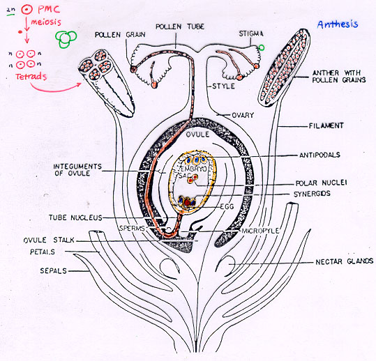
This is the picture where the fertilization takes place.
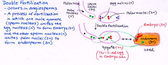
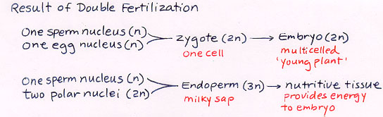
Both of this picture show what is the meaning or what you have to know in double fertilization.
No comments:
Post a Comment